-
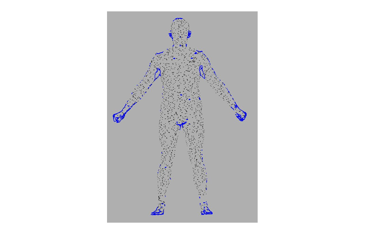
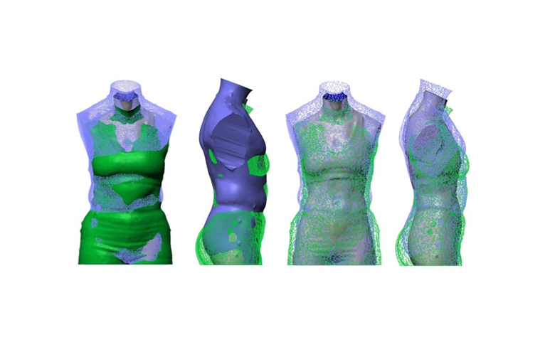
 Figure 6.1Whole body scan showing areas of missing data in blue.
 -
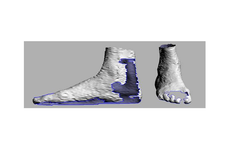
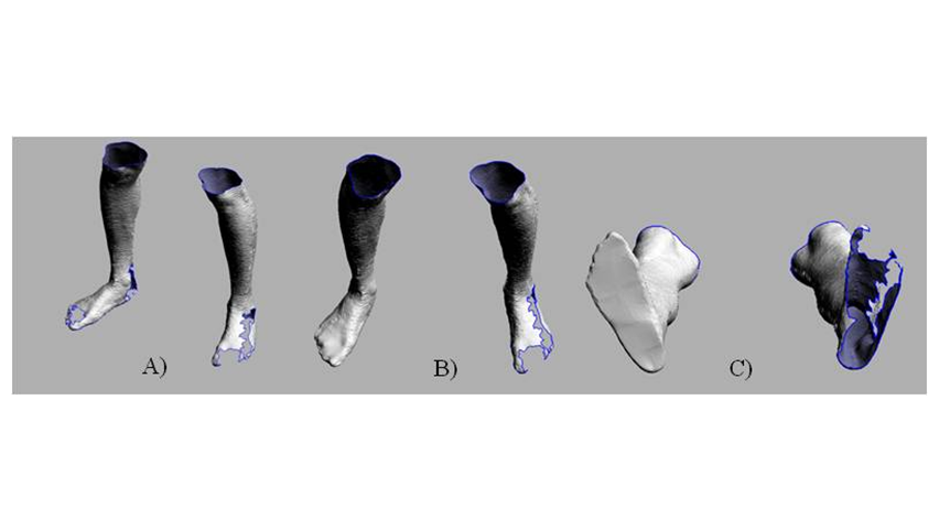
 Figure 6.2Feet segmented from a 3D whole body scan showing holes in scan image.
 -
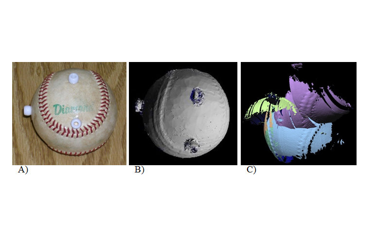
 Figure 6.3(A) Photo of ball. (B) Scan of ball with no metal around. (C) Scan of ball with metal 10 ft from receiver and transmitter.
 -
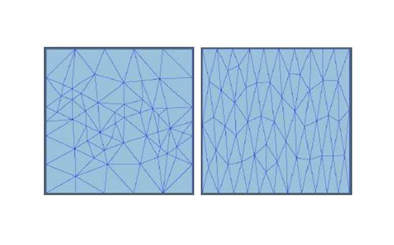
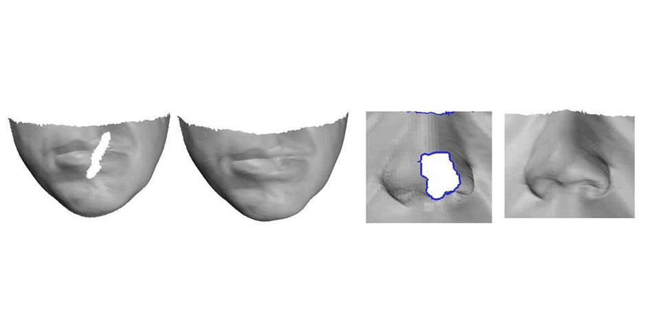
 Figure 6.4Original mesh (left) and smoothed mesh (right).
 -
 Figure 6.5Comparison of filled and unfilled feet from a whole body scan, (A) unfilled holes. Holes filled in one foot, (B) dorsal view and (C) plantar view.
 -
 Figure 6.6Examples of a hole filling algorithm using Hermite splines.
 -
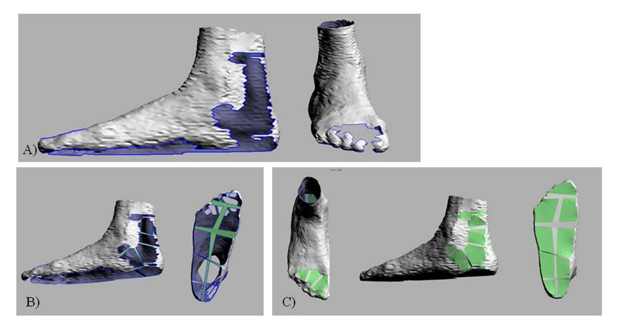
 Figure 6.7Filling holes in feet from a whole body scan (with CySlice 2.0). (A) Unedited scan. (B) Bridging large gaps to approximate foot surface (green). (C) Reconstructed surface (green).
 -
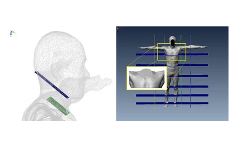
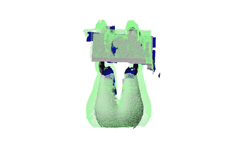
 Figure 6.8Segmenting the whole body scan image
 -
 Figure 6.9Superimposition of multiple subjects
 -
 Figure 6.103D Scan overlay of multiple subjects to determine the overall contour of the seat pan
 -
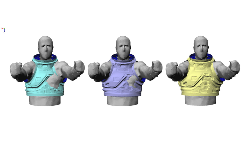
 Figure 6.11Merging of CAD Models with the 3D scan Images
 -
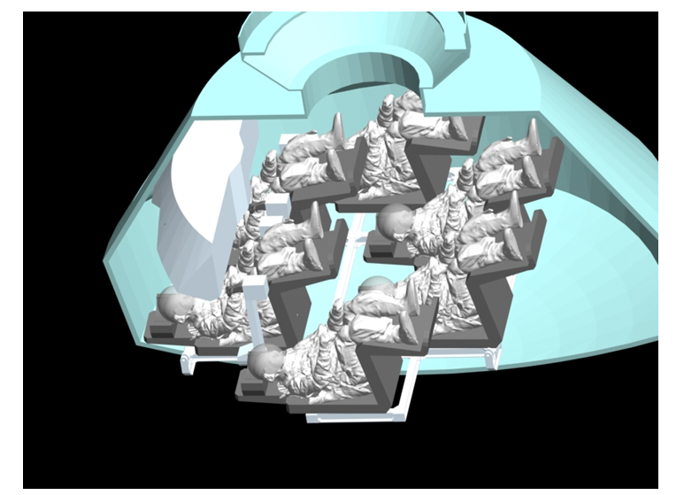
 Figure 6.12Suited crew in a seated posture with 3D Scan data.