-
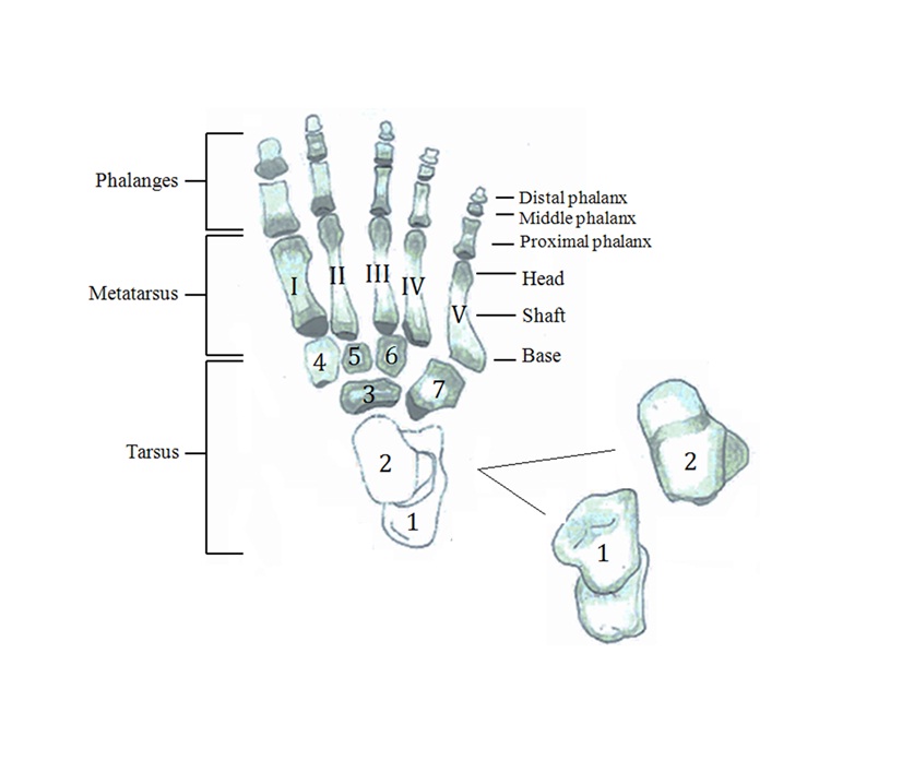
 Figure 1.1Top view of the skeletal foot. Note: 1 calcaneus; 2 talus; 3 navicular; 4 the medial cuneiform, 5 the intermediate cuneiform; 6 the lateral cuneiform; 7 cuboid
 -
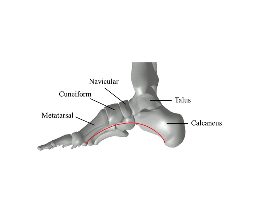
 Figure 1.2The medial longitudinal arch
 -
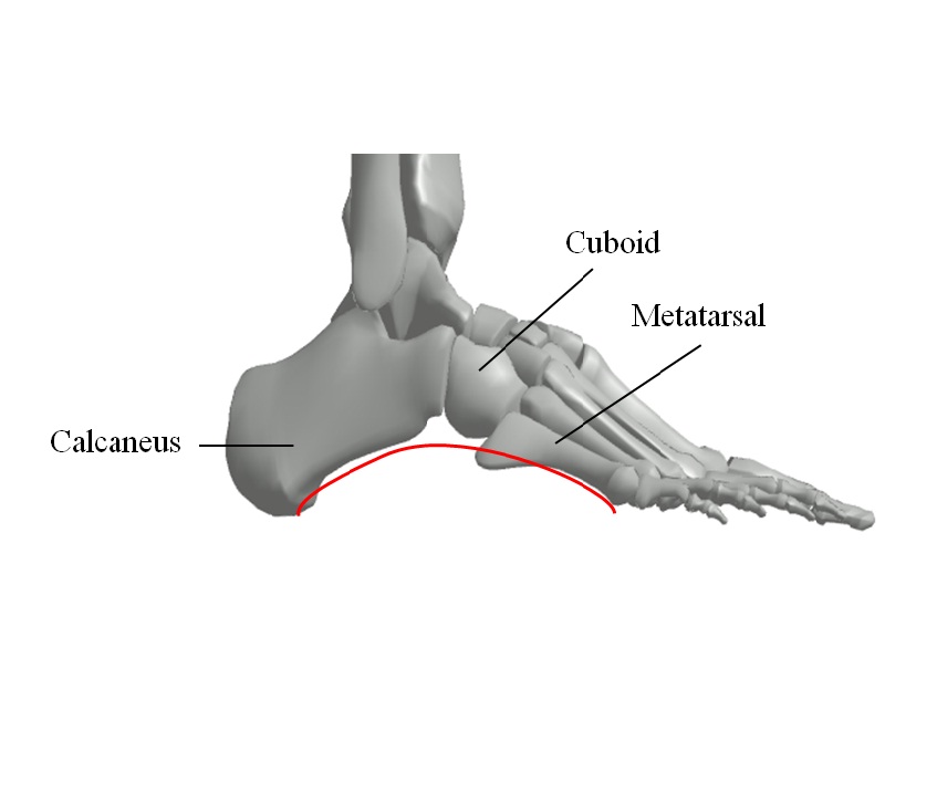
 Figure 1.3The lateral longitudinal arch
 -
 Figure 1.4Transverse arch
 -

-
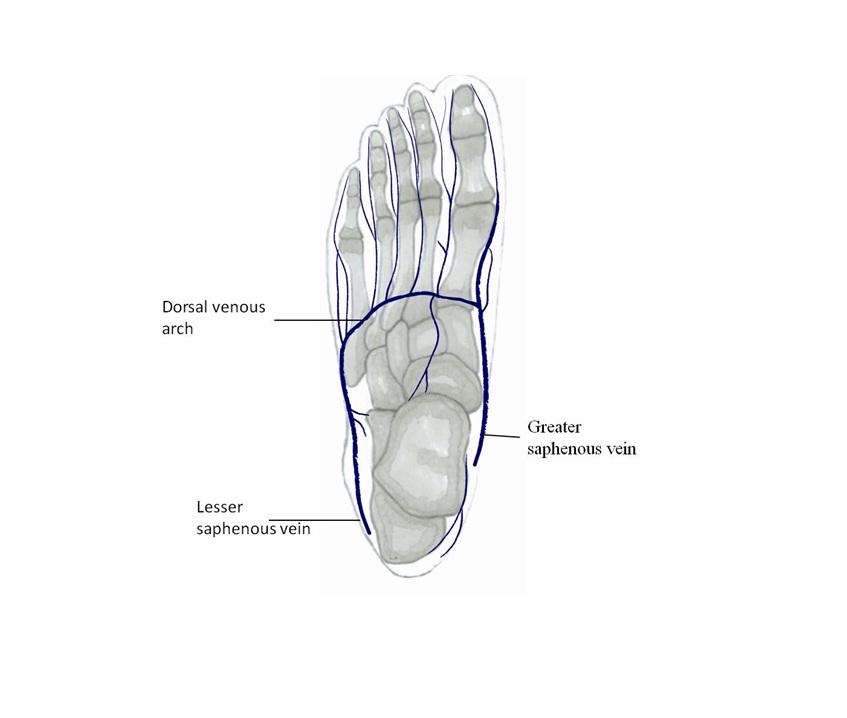
 Figure 1.7The dorsal vein distribution
 -
 Figure 1.8The dorsal vein distribution
 -
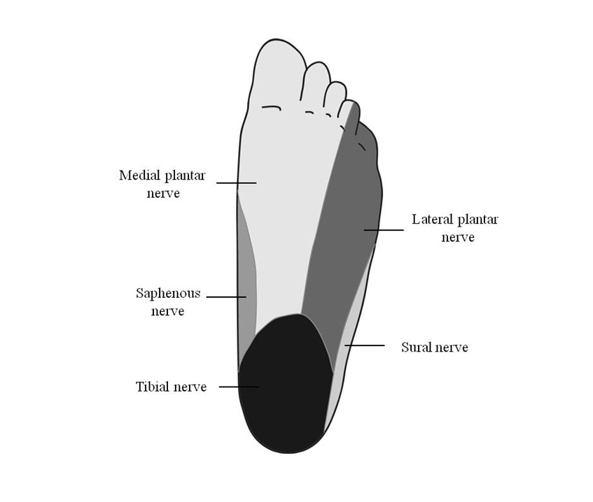
 Figure 1.9The plantar cutaneous distribution
 -
 Figure 1.10The plantar cutaneous distribution
 -
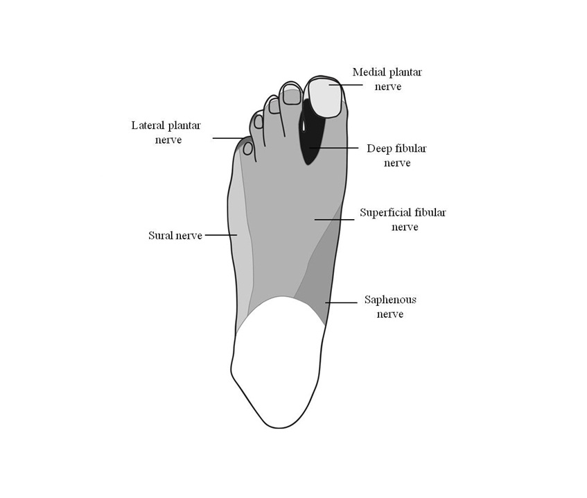
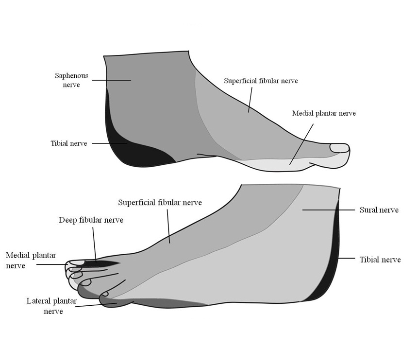
 Figure 1.10The cutaneous distribution on the medial side (above) and lateral side (below)
 -
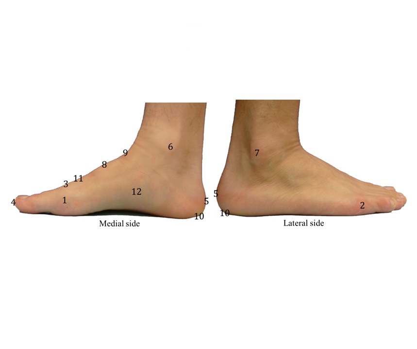
 Figure 1.12Landmarks on the surface of the foot
 -
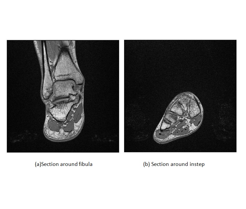
 Figure 1.13Example of MRI images
 -
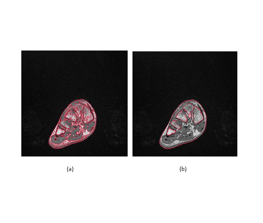
 Figure 1.14Edges extraction from MRI images (a) Automatic edge detection using Matlab program (b) Selection of only the edges of the bones
 -
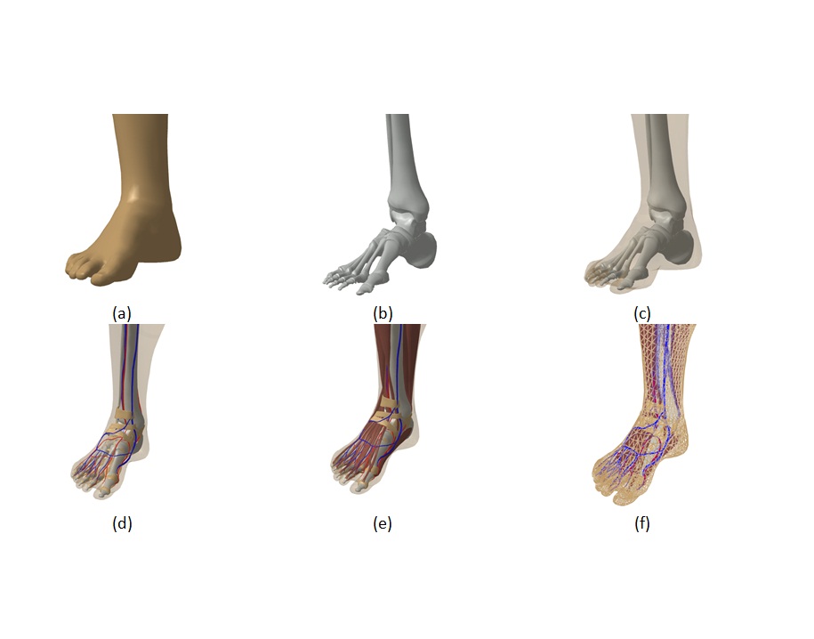
 Figure 1.15Digital model of foot (a) Surface model (b) Skeletal model (c) Visualization of bones and surface (d) Blood vessels and connective tissues (e) Muscles model (f) Mesh model